The starting of life: from fertilisation to blastocyst
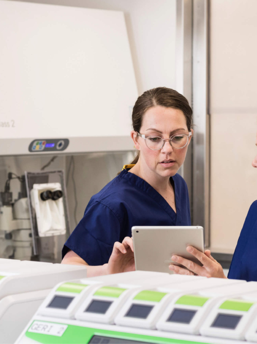
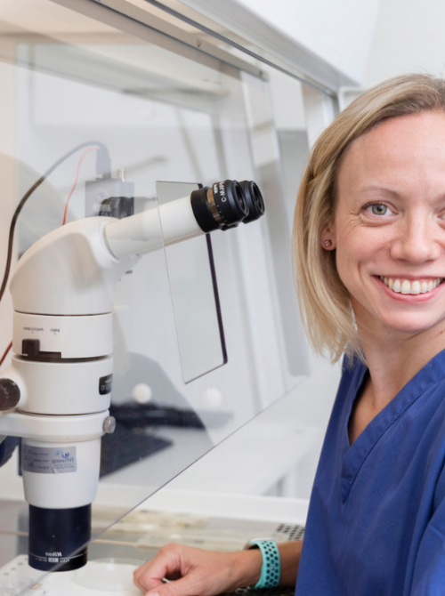
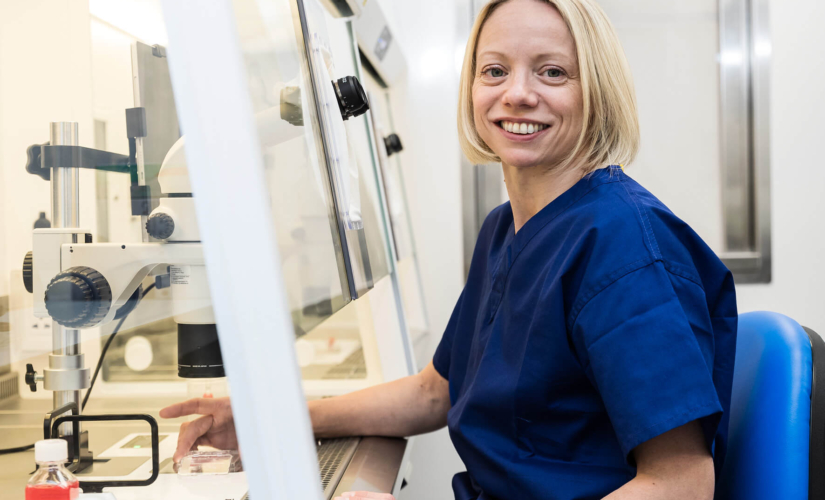
Millie Kanani, Lab Manager at The Evewell Harley Street, explains the fertilisation to blastocyst development in an IVF cycle.
The journey of human life is a marvel of biological complexity, beginning with a single, microscopic cell that has the potential to develop into a fully formed human being.
In the context of IVF, this journey is carefully orchestrated by embryologists, within a controlled environment, involving meticulous scientific techniques and a deep understanding of early human development. While the basic biological processes mirror those of natural conception as much as a lab can permit, the journey in IVF is one of precision, medical intervention, and careful monitoring to ensure the best possible outcomes.
We asked Millie Kanani, our lab manager at The Evewell Harley Street, to explain the process of fertilisation and blastocyst development.
Fertilisation: creating the embryo in the lab
Unlike natural conception, where fertilisation occurs within the fallopian tubes, in IVF or ICSI, this critical event is coordinated by highly skilled and trained embryologists in a lab.
There are two primary methods used for fertilisation in fertility treatment:
- IVF – The eggs collected earlier are ‘inseminated’ with a specific number of motile sperm, in other words, the eggs and sperm are placed together in a culture dish and this allows for the sperm to swim and penetrate the egg – much like it would in natural conception.
- Intracytoplasmic sperm injection (ICSI) – ICSI is most often used when sperm quality is a concern or in cases of previous IVF failures. In ICSI, a single sperm is carefully injected directly into the cytoplasm of the egg using a fine needle. This method bypasses some of the biological barriers to fertilisation, especially in cases of male infertility.
The first few hours…
At fertilisation, the egg undergoes a series of rapid changes, much like in natural conception. If the eggs have been inseminated with sperm via IVF, the sperm penetrates the outer layer of the egg, triggering the cortical reaction, which prevents any additional sperm from entering.
Over the next few hours, the genetic material from the sperm and egg begin to combine, forming the first visual signs that the egg is now… an embryo. This embryo, or ‘zygote’ will have a unique set of 46 chromosomes – the genetic blueprint for a new human being.
At this stage, the embryologist checks to see whether they can see fertilisation has occurred!
A chromosomally normal embryo is called a euploid embryo, whereas a chromosomally abnormal embryo is referred to as aneuploid. We won’t know at this stage if the fertilised egg is normal, or abnormal, we can only test for this after it reaches the blastocyst stage (day 5).
Close monitoring
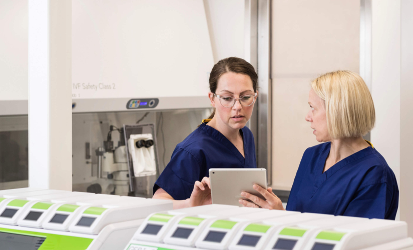
Used as standard in The Evewell labs, timelapse incubators keep the fertilised eggs at body temperature, continually monitoring their development without having to remove them and therefore giving your embryos the best possible chance.
This consistent environment means our lab team can observe and evaluate your embryo development and use this information to potentially influence the selection of embryos for transfer, freezing and thawing.
Cleavage: the early stages of embryo development
Following successful fertilisation, the zygote begins to divide, in a process known as ‘cleavage’. The zygote divides into two cells, then four, eight, and so on, without increasing in overall size. These early divisions occur rapidly, with the embryo typically reaching the eight-cell stage by day three.
As the cells continue to divide, they also begin to compact, a process known as ‘compaction’, where the cells adhere tightly to one another, forming a morula, derived from the Latin word for mulberry – as the embryo at this stage looks like a berry!
This stage is crucial as it signals the beginning of differentiation – the cells are no longer identical and begin to take on specific roles in the embryo’s development.
And this is where timelapse incubators are so vital to our role, as we’re able to watch this process sped up and see if there are any irregular patterns, possibly indicating the first signs of successful blastocyst development, or not.
Blastocyst formation: a key milestone
By day five or six after fertilisation, the embryo reaches a critical stage known as the ‘blastocyst’ stage. This stage is a significant milestone in IVF, as it is often the point at which the embryo’s viability is assessed for transfer back into the uterus.
The blastocyst consists of an outer layer of cells called the ‘trophectoderm’, which will eventually form the placenta and an ‘inner cell mass’ that will develop into the foetus.
The formation of the blastocyst also involves the creation of a fluid-filled cavity called the ‘blastocoel’. As the blastocyst develops, it sheds its outer shell, known as the ‘zona pellucida’, in a process called ‘hatching’. This is essential for the embryo to be able to implant into the uterine lining.
In IVF, not all embryos will reach the blastocyst stage. The ability to use timelapse incubators to monitor and select embryos that successfully reach this stage gives us a significant advantage, allowing us to select the embryos we believe will give you the best possible chance of a successful pregnancy.
Embryo transfer: the culmination of IVF
Once the embryos have reached the blastocyst stage, the next step in IVF is the embryo transfer. The timing of the transfer is critical and is usually done five days after fertilisation, although in some cases, embryos may be transferred earlier.
We work hard to give our patients options and choices, minimising pregnancy loss and creating families – both now and in the future. Your consultant will plan as much as possible into the pre-transfer stage to maximise the chances of success, for example: blood tests to check hormone levels, gynaecological investigations and considering adding PGT-A into your treatment plan.
If you have plans for more than one child, we may discuss with you the choice of more than one egg collection cycle before considering an embryo transfer, to create options for the future. And if this is the case, you will most probably have a frozen embryo transfer further down the line.
Implantation: the beginning of pregnancy
After embryo transfer, the final and perhaps most crucial step is implantation. If successful, the embryo will attach to the uterine lining, a process that typically occurs 6 to 10 days after fertilisation (therefore in a typical IVF cycle, 1-5 days after embryo transfer).
Once implanted, the embryo begins to secrete hormones, including human chorionic gonadotropin (hCG), which signals the body to maintain the uterine lining and support the pregnancy.
In IVF, the success of implantation is not guaranteed, and this period between embryo transfer and pregnancy test (known as the ‘two-week wait’ – although in reality, it’s actually around 10 days!) is often the hardest stage of the IVF process.
Some patients may feel the embryo implantation as a sharp pain, some may experience implantation bleeding, and a lot of patients don’t feel a thing. In the first few days at least, symptoms are usually associated with the side effects of any ovarian stimulation or ovulation medication you’ve been on, or continue to be on – for example, progesterone.
Each patient experiences the process of embryo implantation differently. Some women don’t experience any symptoms, and others will notice every little change in their body.
Blood tests to measure hCG levels are typically performed between 10-14 days after the transfer to confirm whether implantation has occurred and a pregnancy has begun.


